
Luminescent rare-earth-based inorganic nanoparticles (nanophosphors) are promising bioimaging agents due to their high photostability, sharp emission bands and relatively low toxicity (Escudero et al, 2016a). Flame aerosol technology provides a scalable (Mueller et al, 2003) and highly reproducible (Strobel & Pratsinis, 2007) process for production of such nanophosphors (e.g. Y2O3:Eu3+,Tb3+) with precise control of their composition and properties (Sotiriou et al, 2011; Sotiriou et al, 2012). Nanophosphors that can be excited in the near-ultraviolet and visible region, such as YVO4:Eu3+,Bi3+ (Takeshita et al, 2008), provide a useful tool for bioimaging (Escudero et al, 2016b) and in vitro dosimetry (Halamoda-Kenzaoui et al, 2015) studies using conventional fluorescence microscopes.
Here, YVO4:Eu3+,Bi3+ nanophosphors are made by flame spray pyrolysis. The optimal Bi content for maximum red-shift of their excitation band edge towards the visible region is identified through systematic experiments. The nanophosphors with the optimal composition are highly crystalline and appear bright red under a conventional fluorescence microscope. Their photostability during dynamic imaging of HeLa cells in vitro is confirmed, contrary to commercial fluorescent (organic-dye labeled) SiO2 nanoparticles that exhibit 50% photobleaching within 3.5 h (Fig. 1).
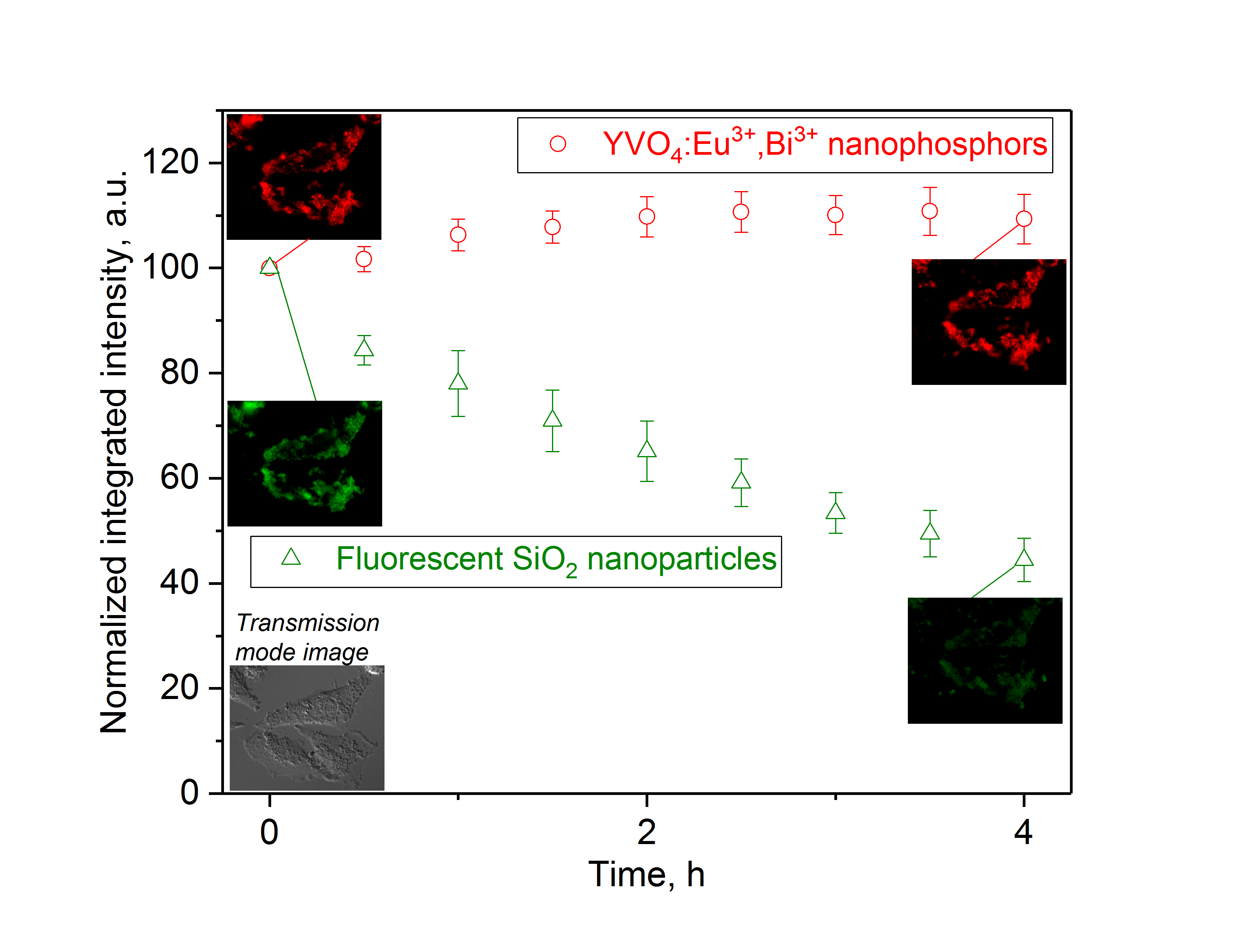
Figure 1. Dynamic fluorescence microscopy imaging of HeLa cells co-incubated with YVO4:Eu3+,Bi3+ nanophosphors and fluorescent SiO2 nanoparticles. The fluorescence intensity of images corresponding to YVO4:Eu3+,Bi3+ is stable with time (circles and red images), while that for fluorescent SiO2 decreases down to 50% within 3.5 h (triangles and green images). The corresponding optical microscopy image in transmission mode at 0 h is also shown.
Furthermore, the deposition rate of these nanophosphors is measured by optical absorption spectroscopy, indicating slower deposition rate in serum-containing than serum-free cell culture medium, consistent with previous studies for different nanomaterials (Allouni et al, 2009; Spyrogianni et al, 2016). This is also confirmed in vitro by monitoring the fluorescence intensity of images of HeLa cells after incubation with nanophosphor suspensions in the presence and absence of serum for various time points (Halamoda-Kenzaoui et al, 2015).
Allouni, Z.E., Cimpan, M.R., Hol, P.J., Skodvin, T., & Gjerdet, N.R. (2009) Coll. Surf. B 68, 83-87.
Escudero, A., Carrillo-Carrion, C., Zyuzin, M.V., & Parak, W.J. (2016a) Topics Curr. Chem. 374.
Escudero, A., Carrillo-Carrion, C., Zyuzin, M.V., Ashraf, S., Hartmann, R., Nunez, N.O., Ocana, M., & Parak, W.J. (2016b) Nanoscale 8, 12221-12236.
Halamoda-Kenzaoui, B., Ceridono, M., Colpo, P., Valsesia, A., Urban, P., Ojea-Jimenez, I., Gioria, S., Gilliland, D., Rossi, F., & Kinsner-Ovaskainen, A. (2015) PLOS ONE 10, e0141593.
Mueller, R., Madler, L., & Pratsinis, S.E. (2003) Chem. Eng. Sci. 58, 1969-1976.
Sotiriou, G.A., Schneider, M., & Pratsinis, S.E. (2011) J. Phys. Chem. C 115, 1084-1089.
Sotiriou, G.A., Franco, D., Poulikakos, D., & Ferrari, A. (2012) ACS Nano 6, 3888-3897.
Spyrogianni, A., Herrmann, I.K., Lucas, M.S., Leroux, J.C., & Sotiriou, G.A. (2016) Nanomedicine (Lond.) 11, 2483-2496.
Strobel, R., & Pratsinis, S.E. (2007) J. Mater. Chem. 17, 4743-4756.
Takeshita, S., Isobe, T., & Niikura, S. (2008) J. Lumin. 128, 1515-1522.
See more of this Group/Topical: Nanoscale Science and Engineering Forum