
Introduction:
Heterotopic ossification (HO), the mineralization of soft tissue also known as osseous metaplasia, is often associated with diffusible therapies such as bone morphogenic protein (BMP) used in repair of bone defect1. Maintenance of appropriate osteogenic concentrations with such short half-life diffusible factors at the site of bone repair requires high doses, resulting in a gradient of the bioactive compound in the surrounding soft tissue that can result in ectopic bone formation2-5. In cases where it is associated with joint or spinal tissue, the clinical complications of HO can be serious and costly for health care providers to correct 6-11. Attempts to mediate the delivery of diffusible factors through localized injection and controlled release substrates have met with only limited success as control and localization of the dose remain problematic12-14. Several other studies have examined gene delivery, using viral vectors or infected/transfected stem cells as an alternative to improve localization of osteogenic factors15-18. Direct viral vector delivery as a means of driving osteogenesis remains hampered by imprecise control over which cells are infected and levels of protein expression post infection. Injection of infected/transfected cells provides an effective means of localization but safety concerns related to levels of osteogenic protein expression and temporal control (i.e. when expression is turned on/off) limits enthusiasm for this technique.
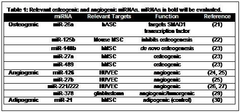
Modulation of gene expression with microRNA (miRNA) is a promising technique for improving control of wound healing and tissue repair processes19. These short, non-coding RNA's are involved in post-transcriptional regulation pathways and in their mature form are found as single strands often associated with RNA silencing complexes20. MicroRNA's utilized in autologous, adipose derived mesenchymal stem cells (ASC) are an attractive therapeutic modality for regenerative medicine. Directing the osteogenic and angiogenic differentiation of ASC is of particular interest in the design of therapies for critical size bone defects, spinal fusion and skeletal reconstruction. A summary of relevant osteogenic and angiogenic miRNA sequences can be found in Table 1. Four miRNAs, miR-26a, -148b, -27a and -489, have shown to regulate osteogenesis in mesenchymal stem cells (MSC). Of these compounds, miR-148b has been shown to induce de novo osteogenesis as indicated by increases in total alkaline phosphatase activity and early specific biomarker expression23. Several miRNAs, miR-126, -27b, -221, -222, -378 and let-7f, play a role in the regulation of angiogensis and tumorigenesis (Table 1). While the expression of all six of these miRNA's modulate angiogenesis, none have demonstrated de novo angiogenic activity and only miR-126 has been explored in stem cells24,25.
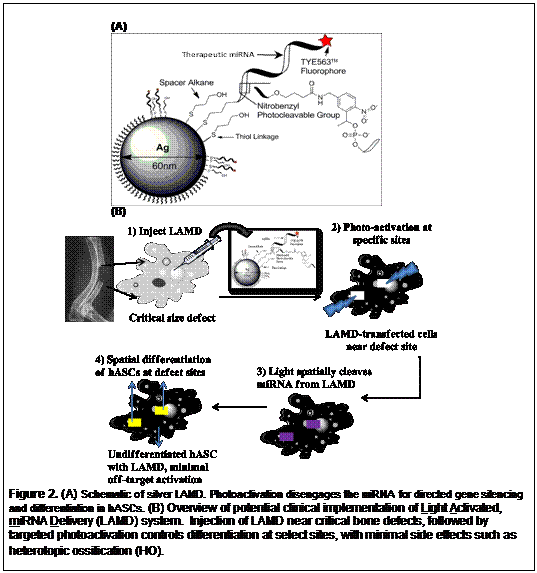
The Light Activated miRNA Delivery (LAMD) system can serve as a potential solution to HO and related complications by providing greater temporal and spatial control over the activation of differentiation (Figure 1). While the LAMD or cells transfected with LAMD may diffuse in vivo, they remain inert until activated with light, providing simple spatial regulation. Additionally, since miRNA are short lived and only transiently modulate mRNA expression, it is expected that there is little chance of permanent activity once an activated cell has begun differentiation. The miRNA associated with inactivated LAMD are expected to degrade slowly intracellularly or in the intercellular space through enzymatic action or hydrolysis. This represents a paradigm shift from current methods of delivering osteogenic compounds such as diffusible BMP or gene delivery for BMP where there is little spatial or temporal control of expression respectively. The LAMD platform addresses both of these issues as: 1) miRNA payload can be activated with focused laser precision allowing activity only in the area of the bone defect, and 2) because miRNA are labile, activity is transient resulting in pathway regulation only in the critical early stages of cell differentiation. Additionally, as the miRNA will only be transfected efficiently when bound to the particle, miRNA from LAMD activated in the intercellular space will have little impact on surrounding tissue.
Materials and Methods:
LAMD synthesis and characterization. Gold and silver based LAMD will be synthesized and characterized by a modified Lee and Meisel method31. As the LAMD shelf-life is unknown, this activity will span the entire period of the project. The particle morphology and degree of functionalization will be characterized by a combination of electron microscopy (EM), dynamic light scattering and zeta potential (DLS), inductively couple plasma optical emission spectroscopy (ICP-OES), gel electrophoresis, UV-Vis, fluorescence spectroscopy, and thermogravimetric analysis. The size of NP used in this study has been informed by previous experimental data that describes maximum field enhancement, nanoparticle uptake and minimum cytotoxicity in the 40-60nm diameter range32-36.
In vitro Photoconversion Efficiency. Photorelease of oligonucleotides from LAMD will be achieved with exposure to 308nm via standard gel transilluminator (100W), 365nm via a mercury arc lamp (206 mW/cm2), 488nm via nitrogen laser or 1064nm laser (Lasermate continuous Nd:YAG laser). Extinction coefficients for the internal, photocleavable 1-(2-nitrophenyl)ethyl (NPE) linker will be calculated according to UV-VIS spectra. Released oligonucleotides in solution are quantified via fluorometry and fluorescent gel electrophoresis of the 5'-TYE fluorescent label (solution: 549nm ex/563nm em, Perkin Elmer LSB-50 spectrofluorimeter; gel: (532nm/560LP, Typhoon 8600 scanner). To determine initial oligonucleotide functionalization of particles, dithiothreitol is used to displace the thiol linkages (1% DTT at 37 °C for 15min) followed by centrifugation and fluorescent analysis as above.
In vitro Nuclease Activity. 1ml of prepared oligo conjugate will be resuspended in 500ul of 1x DNase I NEB buffer 4U of DNase I (2U/ml), incubated at 37°C for intervals of 5, 15, and 45min and stopped with 5mM EDTA. Oligo conjugates will be purified via centrifugation and DTT-mediated displacement per above. Free solution and particle conjugates will be desalted via spin columns (3kDa Amicon Ultra filters, Millipore) and quantified for gel fluorescence.
Transfection. Transfection will be conducted according to a modification of previously published protocols19. Treated cells will be incubated for 16 hrs following miRNA addition prior to photoactivation.
In vitro Activity of LAMD. ASC will be incubated for 24hrs in OM, AM or SM media at 37°C prior to LAMD treatment. LAMD, at the appropriate concentration, will be added directly to 8-well chambered slides. Treated cells will be incubated at 37°C in a CO2 incubator for 16 hrs following addition prior to photoactivation (as above). Spatially selective activation will be achieved using a chrome/glass photomask. After photoactivation cells will be returned to culture for 21 days. All samples will be resuspended in Opti-MEM® prior to imaging.
RNA isolation and qRT-PCR. This method is previously described by Kilroy et al. 37. Samples will be isolated from LAMD treated ASC at 7,14 and 21 days. Primers for angiogenic markers CD31 (PECAM), CD34, CD144 (VE-cadherin), and endothelial cell nitric oxide synthase (eNOS) have been previously described by Cao et al.38. Primers for osteogenic markers alkaline phosphatase (AP) osteocalcin (OCN) have been previously been described by Luzi et al 21. Primers for apidogenic differentiation have been previously described by Yu et al.39.Reactions will be performed with a MJ Mini Thermal Cycler (BioRad). Samples will be normalized (ΔCt) against 18S rRNA.
Flow Cytometery. Flow cytometry will be conducted according to our methods described in Qureshi et al.40
Immunofluorescent Staining & Confocal Microscopy. Indirect immunofluorescent staining and confocal microscopy will be conducted to evaluate LAMD uptake and intracellular fate, miRNA release and expression of differentiation markers as previously described 41. CD31 and von Willebrand's factor will be used as angiogenic markers. AP, OCN and osteopontin (OP) will be used as osteogenic markers and Oil Red O will be used to indicate lipid accumulation and adipogenesis.
Statistical Analysis. All results will be expressed as mean ± SEM. Normality will be confirmed using the Shapiro-Wilk test (P < 0.001). Data will be analyzed with one-way analysis of variance, followed by Tukey's minimum significant difference post hoc test for pairwise comparisons of main effects. A P-value of 0.05 will be considered significant.
Preliminary Results: Directed Differentiation of hASC with miRNA
To evaluate the ability of miR-148b to modulate osteogenesis, hASCs in stromal control media (SM) or osteogenic media (OM) were transfected with increasing doses of RNA oligonucleotides mir-148b, mir-148b* or hybridized miRNA (mir-148b+mir-148b*) for 48 hours. At day 6 and 21 post transfection mRNA expression of the early stage and middle stage osteogenic differntiation marker, alkaline phosphatse (ALP) and osteocalcin (OCN) were measured by semi-quantative PCR, respectively, normalized to 18s RNA (n=3 for each concentration).
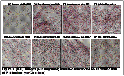
Expression of ALP was examined qualitatively by staining miRNA transfected cells with a colorimetric ALP enzymatic detection kit (Chemicon). hASC cells in stromal control media (SM) and osteogenic media (OM) were transfected with 400nmol of mir-148b, mir-148b* and hybridized miRNA (mir-148b+mir-148b*), previously detemined to be the best for ALP mRNA expression, for 48 hours and stained live on day 6. A substantial difference in color change as a result of dye substrate conversion by ALP can be seen in miRNA treated cells compared to SM control cultures, with a similar but less pronounced difference seen in OM control (Figure 2). These results indicate that increased ALP mRNA expression correlates with an increase in ALP protein production.
References:
1. McCarthy E, Sundaram M. Heterotopic ossification: a review. Skeletal radiology. 2005;34(10):609-19.
2. Harel D. High-dose bone morphogenetic protein‚ induced ectopic abdomen bone growth. The Spine Journal. 2010;10(2):e1-e4.
6. Riegler HF, Harris CM. Heterotopic bone formation after total hip arthroplasty. Clinical Orthopaedics and Related Research. 1976;117:209.
12. Shields LBE, Raque GH, Glassman SD, Campbell M, Vitaz T, Harpring J, et al. Adverse effects associated with high-dose recombinant human bone morphogenetic protein-2 use in anterior cervical spine fusion. Spine. 2006;31(5):542.
15. Kirker-Head CA. Potential applications and delivery strategies for bone morphogenetic proteins. Advanced Drug Delivery Reviews. 2000;43(1):65-92.
19. Rhim C, Cheng C, Kraus W, Truskey G. Effect of MicroRNA Modulation on Bioartificial Muscle Function. Tissue Engineering Part A. 2010:3589-97.
20. Ambros V. The functions of animal microRNAs. Nature. 2004;431(7006):350-5.
21. Luzi E, Marini F, Sala SC, Tognarini I, Galli G, Brandi ML. Osteogenic differentiation of human adipose tissue-derived stem cells is modulated by the miR-26a targeting of the SMAD1 transcription factor. Journal of Bone and Mineral Research. 2008;23(2):287-95.
23. Schoolmeesters A, Eklund T, Leake D, Vermeulen A, Smith Q, Aldred SF, et al. Functional Profiling Reveals Critical Role for miRNA in Differentiation of Human Mesenchymal Stem Cells. PLoS One. 2009;4(5):9.
24. Fish JE, Santoro MM, Morton SU, Yu S, Yeh RF, Wythe JD, et al. miR-126 regulates angiogenic signaling and vascular integrity. Developmental cell. 2008;15(2):272-84.
26. Kuehbacher A, Urbich C, Zeiher A, Dimmeler S. Role of Dicer and Drosha for endothelial microRNA expression and angiogenesis. Circulation Research. 2007;101(14956205421449113292related:zO4-5ZYdj88J):59.
29. Lee DY, Deng Z, Wang C-H, Yang BB. MicroRNA-378 promotes cell survival, tumor growth, and angiogenesis by targeting SuFu and Fus-1 expression. Proceedings of the National Academy of Sciences. 2007;104(51):20350-5.
30. Barhoumi A, Huschka R, Bardhan R, Knight MW, Halas NJ. Light-induced release of DNA from plasmon-resonant nanoparticles: Towards light-controlled gene therapy. Chemical Physics Letters. 2009;482(4-6):171-9.
31. Lee PC, Meisel D. ADSORPTION AND SURFACE-ENHANCED RAMAN OF DYES ON SILVER AND GOLD SOLS. Journal of Physical Chemistry. 1982;86(17):3391-5.
32. Chithrani BD, Ghazani AA, Chan WCW. Determining the size and shape dependence of gold nanoparticle uptake into mammalian cells. Nano Letters. 2006;6(4):662-8.
37. Kilroy GE, Foster SJ, Wu X, Ruiz J, Sherwood S, Heifetz A, et al. Cytokine profile of human adipose-derived stem cells: expression of angiogenic, hematopoietic, and pro-inflammatory factors. J Cell Physiol. 2007;212(3):702-9.
38. Cao Y, Sun Z, Liao L, Meng Y, Han Q, Zhao RC. Human adipose tissue-derived stem cells differentiate into endothelial cells in vitro and improve postnatal neovascularization in vivo. Biochemical and Biophysical Research Communications. 2005;332(2):370-9.
39. Yu G, Wu X, Dietrich MA, Polk P, Scott LK, Ptitsyn AA, et al. Yield and characterization of subcutaneous human adipose-derived stem cells by flow cytometric and adipogenic mRNA analyzes. Cytotherapy. 2010;12(4):538-46.
40. Qureshi AT, Monroe WT, Lopez MJ, Janes ME, Dasa V, Park S, et al. Biocompatible/bioabsorbable silver nanocomposite coatings. Journal of Applied Polymer Science. 2011;120(5):3042-53.
41. Nakajima N, Takahashi T, Kitamura R, Isodono K, Asada S, Ueyama T, et al. MicroRNA-1 facilitates skeletal myogenic differentiation without affecting osteoblastic and adipogenic differentiation. Biochemical and Biophysical Research Communications. 2006;350(4):1006-12.
See more of this Group/Topical: 6th International Conference on Bioengineering and Nanotechnology