
Introduction: Small-diameter synthetic vascular grafts have high failure rate due to thrombogenic responses. In addition, the lack of endothelialization in synthetic grafts results in low patency rate in the long-term. In the past decade, significant progress has been made to construct tissue-engineered blood vessels (TEBV) in vitro by using vascular cells with or without scaffolds. However, constructing cellular grafts involves extensive manipulation of cells in vitro and is time consuming, which limits the application to individualized and non-urgent therapies. Stromal cell–derived factor-1α (SDF-1α) is a potent factor for endothelial progenitor cell (EPC) homing as well as neovascularization. In this study, we investigated the effects of immobilized SDF-1α on the recruitment of EPCs and smooth muscle progenitor cells (SMPCs) to vascular grafts. We propose an in situ tissue engineering approach that recruits two types of endogenous progenitor cells for the regeneration of blood vessels.
Materials and Methods: Microfibrous vascular grafts were fabricated by electrospinning poly-L-lactide (PLLA) and poly-caprolactone (PCL) polymer blends onto a rotating mandrel. Heparin was conjugated to microfibrous vascular grafts to suppress thrombogenic responses, and SDF-1α was immobilized onto heparin to recruit endogenous progenitor cells. To evaluate the effects of surface modification on the patency of vascular grafts in vivo, untreated grafts, heparin-treated grafts and heparin-SDF-1α-treated grafts with a length of 6 mm and an inner diameter of 1 mm were implanted into the left common carotid artery of rats by anastomosis and examined at various time points for up to 12 weeks. Untreated and heparin-treated grafts were used as controls for comparison to determine the effect of immobilized SDF-1α.
Results and Discussion: Heparin-bound SDF-1α was more stable than adsorbed SDF-1α under both static and flow conditions. ELISA measurements showed that SDF-1α was stably immobilized on the microfibrous scaffolds and a slow release of SDF-1α was achieved for more than a week in vitro. Heparin coating improved the short-term patency, and the long-term patency was further improved by immobilized SDF-1α. Within 1 week, SDF-1α effectively recruited EPCs to the luminal surface of the grafts (Figure 1 A-C), without significant effect on the recruitment of inflammatory cells. EPCs differentiated into endothelial cells (ECs), resulting in a complete coverage of luminal surface within 4 weeks. In contrast, the grafts without SDF-1α showed much slower endothelialization, especially in the middle portion. In addition, SDF-1α increased the recruitment of SMPCs to the outer surface of the grafts at 1 week, and SMPCs differentiated into smooth muscle cells (SMCs) in vivo. Isolated SMPCs expressed stem cell markers and spontaneously differentiated into SMCs in vitro. SDF-1α treated grafts showed more extensive and denser extracellular matrix (collagen-I and elastin) deposition surrounding the vascular grafts than the other two groups. Mechanical tests demonstrated that the elastic modulus of the grafts of all three groups increased significantly over time after implantation. SDF-1α treated grafts possessed significantly higher elastic modulus than the other two groups at 2 weeks, 4 weeks and 12 weeks. To determine the long-term fate of the bioactive vascular grafts and effect of polymer degradation on graft remodeling, heparin-SDF-1α-treated grafts were examined as a representative at 6 months post implantation. Characterization of the middle portion of the grafts showed that the polymer had been partially degraded at certain sites. However, there was no sign of aneurysm formation and mechanical weakening in the wall. The endothelium was maintained intact in the lumen surface of the partially degraded grafts, while some SMCs migrated into the scaffolds and deposited significant amount of elastin. Many microvessels were found in the outer part of the scaffolds indicated that the implanted grafts had been well vascularized and integrated with the surrounding tissue.
Conclusions: This work demonstrates the feasibility of simultaneously recruiting endogenous progenitor cells of ECs and SMCs for in situ blood vessel regeneration. This approach will have broad applications in regenerative medicine.
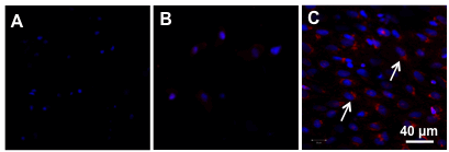
Figure 1. En face immunostaining for CD31 and CD34 of the explanted grafts at 1-week after implantation.
En face immunostaining for endothelial marker CD31 (green) and endothelial progenitor cell (EPC) marker CD34 (red) of the middle portion of untreated grafts (A), heparin-treated grafts (B) and heparin-SDF-1α-treated grafts (C). Arrows indicate CD34+ EPCs. Nuclei were stained by DAPI (blue). Scale bar = 40 μm.
See more of this Group/Topical: 6th International Conference on Bioengineering and Nanotechnology