
Incorporation of Sacrificial Fibers Increases Cell Infiltration and Influences Synovial Stem Cell Behavior
Benjamin Li-Ping Lee1,2, Zhenyu Tang1,2, Fang Huang2, Zhiqiang Yan3, Julia Chu2, Song Li1,2
1 Graduate Program in Bioengineering, University of California at Berkeley and University of California at San Francisco, USA
2 Department of Bioengineering, University of California at Berkeley, Berkeley, CA 94720
3 Institute of Mechanobiology and Medical Engineering, School of Life Science and Biotechnology, Shanghai Jiao Tong University
Tel: 510-666-2799 E-mail: song_li@berkeley.edu
Summary: Neural crest-like synovial stem cells (NCL-SSCs) are isolated, maintained, and characterized. In addition, electrospinning is used to fabricate nanofibrous scaffold containing sacrificial polyglycolic acid (PGA) fibers to improve porosity and enhance cell infiltration. Preliminary studies to assess the effect of increased cell infiltration on NCL-SSCs are presented.
Recent studies have demonstrated that bioactive scaffolds can be used to regenerate cartilage by local cell homing. However, the specific types of cells participating in regeneration are not fully understood. In this study, we identified a novel type of neural crest-like synovial stem cell (NCL-SSC) residing within the synovial membrane, which possesses several important characteristics distinct from previously identified stem cells. Protein marker screening revealed that NCL-SSCs homogeneously expressed neural crest stem cell markers, such as Sox1, Sox10, Snail and Vimentin (Figure 1). In addition, differentiation assays showed that NCL-SSCs can be not only induced into cells of mesenchymal lineages, including osteoblasts, adipocytes, and chondrocytes, but also differentiated into peripheral neurons and Schwann cells. Furthermore, we found that the combination of chick embryo extract and basic fibroblast growth factor can maintain the multipotency and maker expression of NCL-SSCs. Cloning assay showed that the average plating efficiency of NCL-SSCs is about 10% and the cloned colonies retained the marker expression and multipotency. Lastly, NCL-SSCs can form neurosphere-like aggregates when cultured in ultra-low attachment culture dishes.
In addition to an appropriate cell source, tissue engineering often requires a native extracellular matrix-like scaffold. For instance, scaffolds fabricated via electrospinning provide a cell-friendly microenvironment optimal for tissue regeneration. However, one major setback of electrospun scaffolds is the limited cell infiltration due to the small pore size between fibers. Thus, we utilized a dual-composition method to increase the porosity of nanofibrous scaffolds while maintaining the scaffold structure to facilitate cell infiltration. Specifically, poly (L-lactide) (PLLA), a slow-degrading polyester, was used to produce structural nanofibers, and polyglycolic acid (PGA), a relatively fast-degrading polyester, was used to make sacrificial fibers via co-electrospinning. The time-dependent removal of PGA nanofibers via in vitro-degradation resulted in an increase in pore size, a decrease of elastic modulus, and changes in yield strength of the scaffolds (Figure 2). Furthermore, animal studies showed that in vitro-degraded PLLA/PGA scaffolds drastically increased cell infiltration. Most interestingly, although NCL-SSCs seeded on the in vitro-degraded PLLA/PGA scaffolds exhibited similar morphology and proliferation rate when compared with the as-spun PLLA/PGA scaffolds, the enhanced cell infiltration in vitro resulted in a differential regulation on the lineage commitment of NCL-SSCs. In summary, we not only indentified a new and important type of local cells in synovial knee joint, but also incorporated sacrificial PGA fibers to fabricate a porous and functional three-dimensional construct useful for in situ tissue engineering.
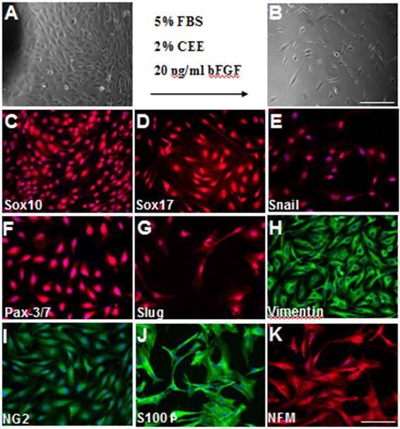
Figure 1. Isolation and characterization of synovial stem cells. Synovial stem cells were cultured in maintenance media with chick embryo extract and basic fibroblast growth factor. (A-B) Synovial stem cells were isolated by using tissue explant culture method and were passaged when confluent. Scale bar is 50 µm. (C-K) The derived synovial stem cells were immunostained with antibodies against Sox10 (C), Sox17 (D), Snail (E), Pax-3/7 (F), Slug (G), Vimentin (H), NG2 (I), S100β (J), and NFM (K). Scale bar is 100 µm.
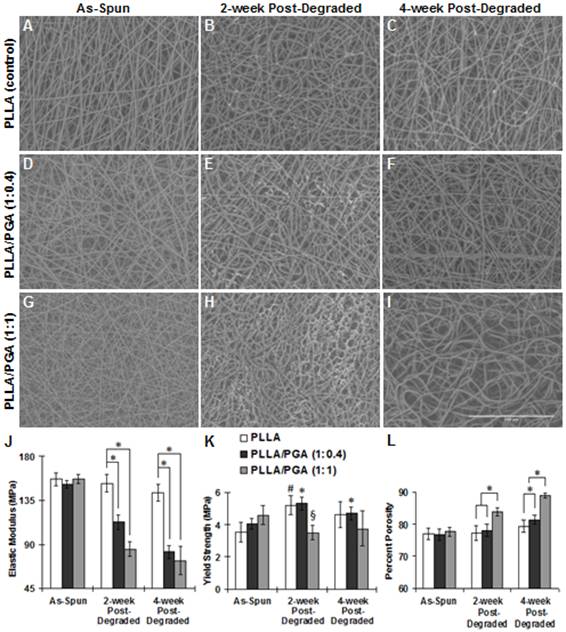
Figure 2. Fibrous structures and mechanical properties of nanofibrous scaffolds with different PLLA/PGA ratio at different time points. SEM was used to visualize the fibrous structures of as-spun, 2-week post-degraded, and 4-week post-degraded scaffolds with pure PLLA (A-C), PLLA/PGA (1:0.4) (D-F), and PLLA/PGA (1:1) (G-I). Scale bar is 100µm. Elastic modulus (J), yield strength (K), and percent porosity (L) were quantified for each scaffold at each time point.
See more of this Group/Topical: 6th International Conference on Bioengineering and Nanotechnology