
Enhanced Islet Survival and Function on Extracellular Matrix Protein-Coated Scaffolds
Woon Teck Yap, David M. Salvay, Michael A. Silliman, Xiaomin Zhang, Zachary G. Bannon, Dixon B. Kaufman, William L. Lowe, Jr. and Lonnie D. Shea
Departments of Biomedical Engineering, Chemical and Biological Engineering, Surgery, and Medicine
Northwestern University, Chicago, IL 60611
Islet transplantation offers the potential to improve glycemic control and reduce insulin dependence in patients with type 1 diabetes mellitus (T1DM), an autoimmune disease that affects over 1.5 million Americans [1]. We have successfully employed biodegradable, microporous scaffolds fabricated from poly(lactide-co-glycolide) (PLG) for islet transplantation [2]. These have been shown to provide a three-dimensional matrix that supports islet attachment following isolation, which resulted in improved islet function and viability. However, the process required to isolate islets severs critical vascular connections and disrupts interactions between the islet cells and their extracellular matrix (ECM). In a previous report, we demonstrated that adsorption of ECM proteins found in the native islet microenvironment to the scaffold surface resulted in enhanced islet function in vivo and significantly decreased the amount of time required to reverse hyperglycemia in a mouse model of T1DM [3]. The current studies extend this work by examining the role of ECM proteins in enhancing islet survival and function, independent from other effects these proteins may mediate in the diabetic recipient, such as promoting host cell infiltration and islet revascularization.
PLG scaffolds were prepared and treated as previously described on the day before islet isolation and seeding [3]. Specific ECM proteins (collagen IV, fibronectin or laminin) were adsorbed to the scaffolds by overnight incubation. Diabetes was induced in C57BL/6 mice receiving islet transplants using streptozotocin. Islet isolation and scaffold seeding were performed as previously described. Islet cell metabolic activity, live/dead cell ratio, glucose-stimulated insulin release and apoptosis were quantified. Histological analyses were also performed to characterize the morphologies and architectures of the transplanted islets, relative to those of islets within freshly-isolated pancreata. Measurements of total islet area, total intra/peri-islet functional vascular area and density were performed in grafts removed 16 or 113 days following implantation. Statistical analyses for comparison of groups were performed by one-way ANOVA with a post-hoc Tukey HSD.
Islets
cultured on protein-adsorbed scaffolds exhibited enhanced metabolic activity
for all conditions tested relative to the control (Figure 1A). The glucose
stimulation indices for islets cultured on collagen IV-adsorbed scaffolds were
significantly higher than the control (Figure 1B). Islets cultured on collagen
IV-adsorbed scaffolds had a significantly higher ratio of live to dead cells and
exhibited significantly reduced levels of caspase-3/7 activation compared to
those cultured on fibronectin- and laminin-adsorbed 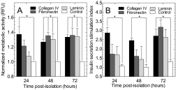 scaffolds
and to the control. Islets seeded onto either the collagen IV-adsorbed
scaffolds or the control scaffolds exhibited morphologies and cellular
architectures that were similar to those in islets within the native stroma of
pancreata. Additionally, the mean total islet area
 per histological section for the
collagen IV group was greater than that for the control group. Interestingly, on
both days 16 and 113 post-transplantation, islets transplanted using both
collagen IV-adsorbed scaffolds and control scaffolds exhibited a dense, perfused
vasculature as visualized by immunofluorescence staining.
Islets cultured on scaffolds adsorbed with collagen IV, fibronectin or laminin displayed increased metabolic activity and insulin secretory capacity, lower levels of cytoxicity, and decreased apoptosis compared to islets cultured on the control scaffolds at all time points. In vivo studies showed that islets transplanted on scaffolds exhibited islet architectures similar to those found in freshly-isolated pancreata. These results indicate that islets seeded onto scaffolds adsorbed with ECM proteins and transplanted into a diabetic mouse model displayed enhanced survival and function.
Based on our in vitro data, we postulate that this effect is due to improved islet metabolism, enhanced insulin secretion and decreased islet cell necrosis and apoptosis, independent from the other effects that these proteins may mediate in the diabetic recipient. These results further establish the use of PLG scaffolds as basic research tools, as well as their utilization in therapeutic applications.
References
[1] Eiselein, L.; Schwartz H.J.; Rutledge J.C. The Ilar Journal. 2004, 45, 231.
[2] Blomeier, H.; Zhang, X.; Rives, C.; Brissova, M.; Hughes, E.; Baker, M.; Powers, A.C.; Kaufman, D.B.; Shea, L.D., Lowe, W.L., Jr. Transplantation. 2006, 82, 452.
[3] Salvay, D.M.; Rives, C.B.; Zhang, X.; Chen, F.; Kaufman, D.B.; Lowe, W.L., Jr.; Shea, L.D. Transplantation. 2008, 1456.
See more of this Group/Topical: 6th International Conference on Bioengineering and Nanotechnology