
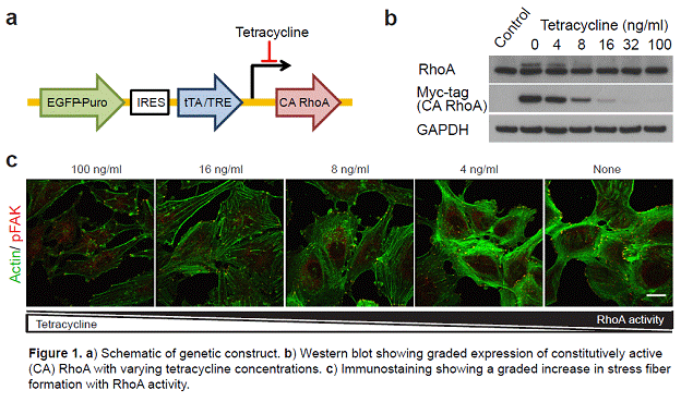
The structural and mechanical properties of cells such as cell size, shape, and stiffness have recently been recognized as important regulators of cell behavior. For example, cells cultured on small areas of adhesive extracellular matrix (ECM) grow slowly and apoptose more frequently than cells spread on large areas (Chen et al., 1997 Science 276: 1425-1428), and cells confined to one-dimensional lines of ECM elongate their cell bodies and can migrate faster than cells cultured on standard unconfined two-dimensional substrates (Doyle et al., 2009 J Cell Biol. 184: 481-490). Others have shown that both ECM patterning and changes in ECM stiffness can alter stem cell self renewal and differentiation. While there are many strategies to indirectly control cell behavior through manipulation of the extracellular environment, there is a marked lack of strategies for controlling these mechanotransductive signaling systems in more direct and precise ways. Such strategies could offer a powerful means with which to control cell behavior at cell-material interfaces. To address this unmet need, we have developed a synthetic biology-inspired strategy for directly tuning the mechanical properties of cells. Specifically, we have placed constitutively active (CA) mutants of two intracellular mechanotransductive signaling proteins: RhoA GTPase and myosin light chain kinase (MLCK) under a tetracycline-repressible promoter and introduced a single copy of these constructs into human glioblastoma cells using viral gene delivery (Fig. 1a). RhoA and MLCK are both known to activate non-muscle myosin II, which is the motor protein responsible for cellular force generation. By expressing these proteins from a tetracycline-repressible promoter, we can vary their activity in a graded fashion by simply changing the concentration of tetracycline in the cell culture medium. This strategy could thus provide a unique opportunity to dynamically alter the mechanical properties and behaviors of cells independently from properties of the ECM.
In this study, we first show that modest expression of CA RhoA from the tetracycline-repressible promoter (only 23% over endogenous RhoA, Fig. 1b) increases the overall activity of RhoA by fivefold. We also evaluate the kinetics of the system by placing a GFP reporter behind the same tetracycline-repressible promoter and measuring intracellular fluorescence following tetracycline withdrawal, which reveals initiation of gene expression within 12 hours and complete expression within 48 hours. Maximal expression of CA RhoA induced by complete withdrawal of tetracycline causes a dramatic increase in the assembly of stress fibers, focal adhesions, and cell-cell adhesions (Fig. 1c). These effects can be modulated in a graded fashion by varying the concentration of tetracycline and are not accompanied by significant changes in expression of key adhesive and motor proteins such as beta-1 integrin, alpha-actinin, vinculin, and myosin II. We then demonstrate that by varying the expression level of either CA RhoA or CA MLCK, we can engineer graded changes in both the cortical stiffness of cells and the traction forces that these cells exert on their surroundings, as measured by atomic and traction force microscopy, respectively. By culturing cells on defined-stiffness matrices, systematically varying tetracycline concentration, and measuring random cell migration, we find that CA RhoA expression inhibits cell migration and that cells are most sensitive to increased RhoA activity when cultured on soft ECMs. Moreover, by reversibly switching expression of CA RhoA on and off over time (by adding and removing tetracycline), we can dynamically control cell spreading, migration, and ECM remodeling. Finally, we show that this strategy offers significantly superior performance to standard pharmacological agents used to activate RhoA; for example, the effects of lysophosphatidic acid are short-lived and the molecule is poorly soluble in water, and the Rac GTPase inhibitor NSC23766 is toxic at the dosages needed to produce clear and sustained effects on cell morphology.
We believe that this genetic strategy for varying the activity of mechanotransductive proteins could serve as a valuable tool for directly controlling how cells physically interact with their microenvironment and could potentially allow one to “re-engineer” how cells respond to ECM properties. In addition, this strategy would be useful for developing quantitative relationships between intracellular signaling pathways, cellular physical properties, and complex cell behaviors. For example, most direct manipulations of mechanotransductive signaling in living cells have focused only on turning specific proteins “on” or “off”, and the relationships describing how cellular properties vary quantitatively with specific signaling proteins is largely unknown. Thus, exploring the effects of more measured changes in the activity of proteins like RhoA and MLCK would advance our quantitative understanding of mechanotransductive signaling and could reveal important nonlinear relationships (such as an optimal level of protein activity for cell migration).
J. L. MacKay, A. J. Keung, and S. Kumar (2012). A genetic strategy for the dynamic and graded control of cell mechanics, motility, and matrix remodeling. Biophysical Journal 102: 434-442
See more of this Group/Topical: 6th International Conference on Bioengineering and Nanotechnology