Lateral Canthopexy: An Anatomical Perspective on the Mechanism of Suture Based Lower Eyelid Support
Methods/Technique: Four fresh cadaveric hemifaces were dissected utilizing 2.5x loupe magnification to define the lateral canthal area, with specific attention to identify the structure of the lateral canthal complex. Lateral canthopexy was then performed by selectively grasping the 1) lateral palpebral raphe (LPR), 2) superficial lateral canthal tendon (SLCT), 3) lateral canthal tendon (LCT), or 4) the lateral edge of the inferior tarsal plate (LEITP) and the excursion of the lower lid margin was assessed and recorded videographically. The insertions of the three components of the lateral canthal complex were then divided. Lateral canthoplasty was then performed by selectively grasping each component of the lateral canthal complex and lower lid excursion was reassessed.
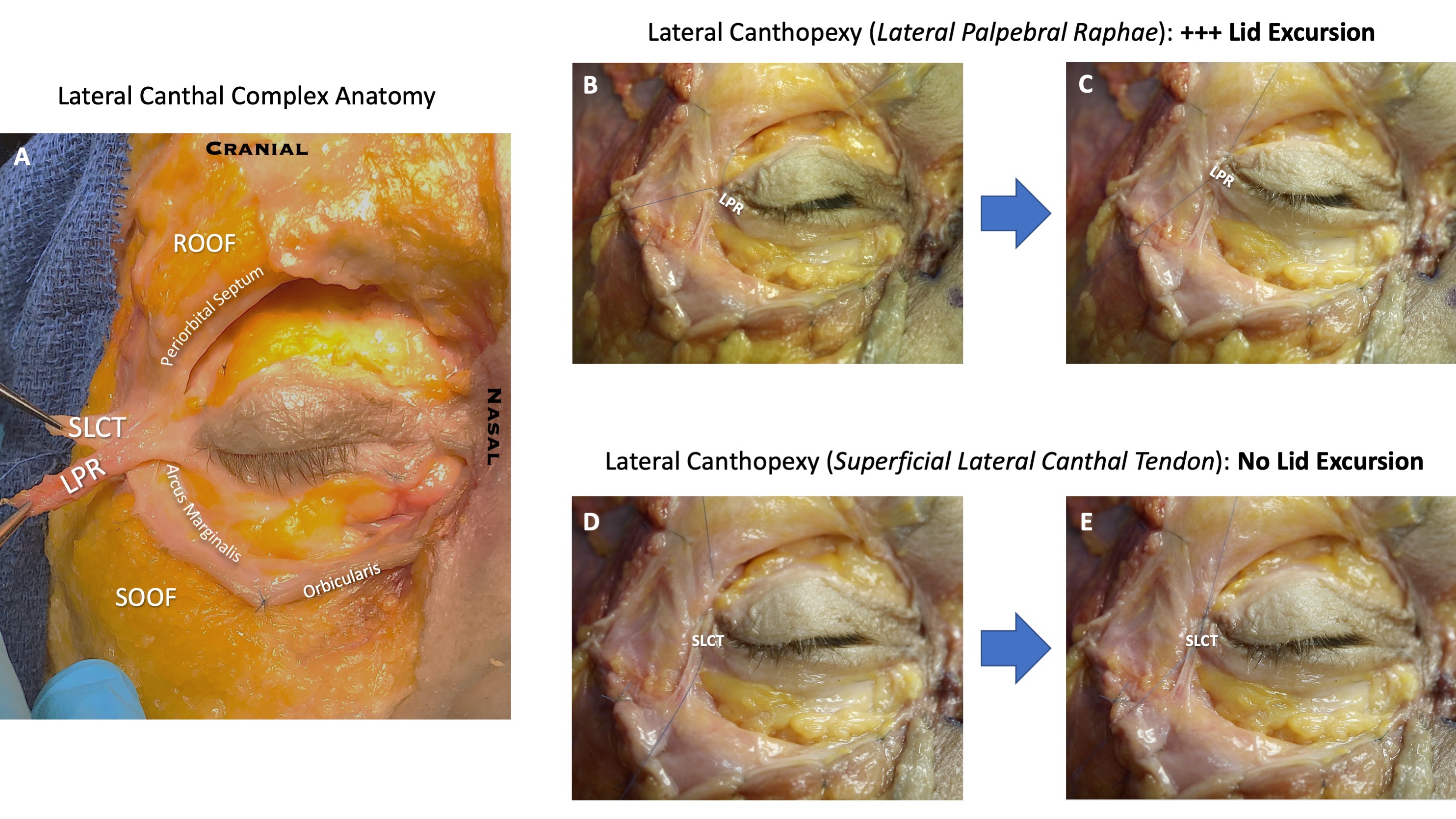
Results/Complications: The three layers composing the lateral canthal complex, which are the LPR, SLCT, and LCT, were confirmed (Figure 1, A). The LPR loosely adherent to the SLCT and was relatively mobile. The SLCT on the other hand was immobile and rigidly fixed to the lateral orbital rim periosteum. The SLCT, also known as the lateral orbital thickening, was continuous with a stout fibrous ring that was fixed to the lateral orbital rim. This continuous fibrous structure continued superiorly as a well-defined strong septum originating from the arcus marginalis forming the inferior boundary of the retro-orbicularis oculi fat (ROOF). It continued inferiorly as the bilamellar orbicularis retaining ligament. Suture fixation of the LPR (Figure 1, B & C) and LEITP resulted in the greatest excursion of the lower lid margin. In contrast, lateral canthopexy utilizing the SLCT resulted in no movement of the lower lid (Figure 1, D & E). Excursion of the lateral canthus via the SLCT required extensive division of the broad SLCT periosteal insertion on the lateral orbital rim along. LCT plication was possible after division and reflection of the SLCT and resulted in minimal excursion of the lower lid.
Conclusion: Based on this anatomic study, the lateral canthal area can be divided into a mobile unit and a fixed unit. For a lateral canthopexy to cause tightening of the lower lid, it should target either the LPR, LEITP or the lateral orbicularis oculi muscle. In contrast, minimal lower lid excursion is observed if the SLCT or LCT are utilized – therefore, lateral canthoplasty is required with extensive release of the retaining ligaments for effective lower lid mobilization utilizing the SLCT or LCT.
Figure 1: Lateral Canthal Complex. A, The Lateral Palpebral Raphae (LPR) is in continuity with the orbicularis muscle and forms the most superficial component of the lateral canthal complex. The Superficial Lateral Canthal Tendon (SLCT) lies deep to the LPR. The SLCT has a broad stout attachment to the lateral orbital rim periosteum and is in continuity with the orbital septum and arcus marginalis. The Lateral Canthal Tendon (LCT) lies deep to the SLCT and inserts ~2mm inside the orbital rim (not shown). The inferior lid orbicularis muscle and orbital septum are reflected inferiorly and secured with suture. B & C, Canthopexy grasping the LPR results in the greatest excursion of the lower eyelid. D & E, Canthopexy grasping the SLCT does not result in lower eyelid excursion.